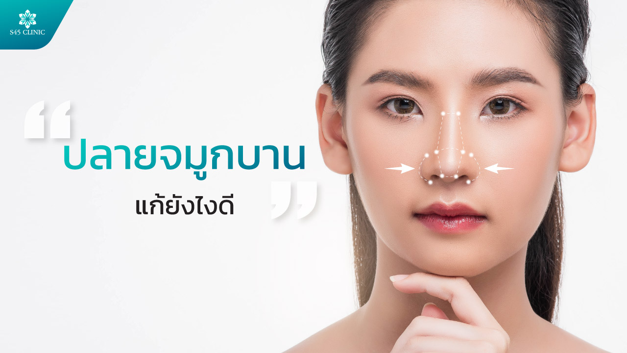
Fascia Graft Rhinoplasty, Can Retroauricular Fascia Correct a Thin Nasal Tip

A thin nasal tip is especially common in people who have had rhinoplasty before. When the tip becomes so thin that the silicone implant shows through, it is a sign of a risk of implant extrusion (the implant pushing through the skin). Using tissue from behind the ear is a revision rhinoplasty method surgeons use to correct a thin nasal tip.
Why Does the Nasal Tip Become Thin
- Genetic causes
- small nasal bones and a low tip structure
- thin skin over the tip from birth
- weak, soft tip cartilage
- little soft tissue around the tip
- Causes from rhinoplasty
- a silicone implant that is too large and presses on the tissue
- a silicone implant in place for a long time, pressing the skin thinner
- an unsuitable surgical technique
- a history of infection or inflammation
- the body rejecting the implant material
Risks of a Thin Nasal Tip
- skin so thin that the silicone implant shows through
- risk of implant extrusion through the skin
- a red nose with visible blood vessels
- an unattractive nose shape that looks artificial
- complications may develop if it is left untreated
What Is Tissue From Behind the Ear
Tissue from behind the ear (conchal cartilage with perichondrium) is ear cartilage together with its covering layer, which surgeons use in revision rhinoplasty. It includes:

- cartilage, which gives structure and strength
- perichondrium (the covering layer), which has a blood supply
- some tissue and fat, which help add volume
Advantages of Tissue From Behind the Ear
- it is your own tissue, which lowers the risk of allergy and infection, as the body is unlikely to reject it
- the strength and softness of cartilage
- it integrates with surrounding structures because it has a blood supply
- the nose shape can be adjusted as planned
Using Tissue From Behind the Ear to Correct a Thin Nasal Tip
1. A layer between the silicone implant and the skin: tissue from behind the ear is placed as a cushioning layer between the implant and the skin, which adds thickness to the skin of the tip, reduces the pressure of the implant on the skin and helps lower the chance of implant extrusion.
Suitable for:
- people who have had silicone rhinoplasty but whose skin is getting thinner from friction
- people who want a higher bridge and a projected tip
2. Replacing the silicone at the tip completely: the surgeon removes the tip part of the silicone implant and uses tissue from behind the ear to build a new tip, possibly together with septal cartilage, to sculpt the desired shape.
Advantages:
- addresses problems caused by the silicone implant, because it is removed
- the nose feels soft to the touch
- no risks from implant material (silicone) in the tip
- the nose shape can be adjusted
3. Strengthening the tip cartilage: the surgeon places tissue to reinforce the structure beside the tip, making the tip stronger and helping prevent the alae from collapsing, which helps the nostrils look balanced on both sides.
- a stronger tip structure
- the nose keeps its shape
Steps Before Revision Rhinoplasty With Tissue From Behind the Ear
- consultation with the surgeon and surgical planning
- physical examination, photos and nose design
- blood tests and a readiness assessment
- no food or drink for 6 to 8 hours before surgery
Surgical Steps
- general anaesthetic, with one-to-one care from an anaesthetist
- harvesting tissue from behind the ear
- a small incision behind the ear
- the cartilage is taken with its covering layer
- about 2cm to 3cm in size
- the wound is closed with fine stitches, and the scar is usually small
- preparing the tissue
- shaping it to the desired form
- it may be carved thinner or layered for more thickness
- designing it to fit the patient’s nose structure
- nose surgery
- the nose is opened through the nostrils or the nasal base
- the old silicone implant is removed (if any) and the tissue cleaned
- tissue from behind the ear is placed in the planned position
- the shape is refined for symmetry
- the wound is closed with dissolvable stitches
Advantages of Using Tissue From Behind the Ear to Correct a Thin Nasal Tip
- a low chance of rejection or allergy, because it is your own tissue
- no artificial material in the graft itself
- a low chance of infection
- strong
- a soft nose that feels like your own
- the shape can be refined in fine detail
- no visible edges or outline of an implant
- more resistant to impact than silicone
- does not usually shrink or dissolve
Scars:
- the incision behind the ear is very small and hidden in the crease
- the nose incisions are inside the nose or at the nasal base, so they are hard to see
- keloid scars are uncommon
Who Is Not Suitable for Revision Rhinoplasty With Tissue From Behind the Ear for a Thin Tip
- people who have had keloid scars on the ear
- people with a bleeding disorder
- people who are pregnant or breastfeeding
- people who have had ear surgery that left too little tissue
- people with unrealistic expectations
If any of these apply to you, tell your doctor so they can give you suitable advice.

Frequently Asked Questions
Is Harvesting Tissue From Behind the Ear Painful
The surgery is done under a general anaesthetic, so you will not feel pain during the procedure. After surgery you may feel slight tightness or numbness for 2 to 3 days, which is usually mild and can be managed with painkillers.
Will the Back of My Ear Change Shape
Usually not, because only a part hidden at the back is taken, which does not affect the visible shape of the ear.
How Long Do I Need off Work
Usually about 7 to 14 days, depending on your type of work and the swelling.
How Much Does Revision Rhinoplasty Cost
It depends on the complexity of the structural problem, the nose shape, the surgeon’s experience, medicines and follow-up care.
Is Tissue From Behind the Ear Hard or Soft
Tissue from behind the ear is much softer than silicone and feels closer to your own nose, but it is firm enough to support the shape.
Note: Repeat nose surgery, which many people know as ‘nose reshaping’, is surgery for people who have had nose surgery before and need a further assessment by a surgeon. The approach to care depends on the original problem, the nose structure and the surgeon’s judgement in each case.